Acta Agronomica Sinica
2018, 44 (
):
483-492.
10.3724/SP.J.1006.2018.00483
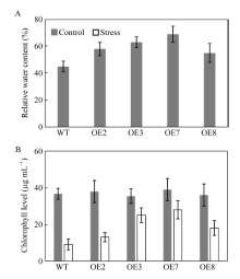
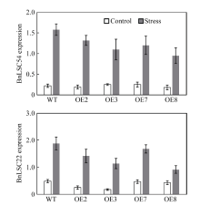
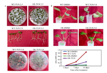
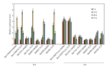
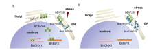
The molecular chaperone binding protein gene participates in the constitutive function of plant growth and protects plant cells against stresses. In this study, we found that BnA7HSP70 overexpressed transgenic lines did not wilt and showed only a small decrease in water potential. However, the wild type lines showed a large decrease in leaf water potential. The transgenic plants had higher relative water content, better osmotic adjustment and less lipid membrane peroxidation. In addition, the leaves from the elevated levels of BnA7HSP70 in transgenic lines conferred tolerance to the glycosylation inhibitor tunicamycin during germination. BnA7HSP70 overexpression-mediated attenuation of stress-induced cell death was confirmed by the decreased percentage of dead cell and the reduced induction of the senescence-associated marker gene BnCNX1. These phenotypes were accompanied by a delay in the induction of the cell death marker genes BnNRP, which are involved in transducing a cell death signal generated by ER stress and osmotic stress through the NRP (N-rich protein)-mediated signaling pathway. Enhanced expression of BnA7HSP70 delayed unfold protein response and NRP pathway mediated chlorosis and the appearance of senescence-associated markers BnLSC222 and BnLSC54 in Brassica napus. These results suggest that overexpression of BnA7HSP70 in Brassica napus alleviate ER stress and osmotic stress-integrating cell death response confronted with water stress.
