Welcome to Acta Agronomica Sinica,

Acta Agronomica Sinica ›› 2021, Vol. 47 ›› Issue (12): 2371-2378.doi: 10.3724/SP.J.1006.2021.01094

• CROP GENETICS & BREEDING·GERMPLASM RESOURCES·MOLECULAR GENETICS • Previous Articles     Next Articles

Virus-mediated expression of GFP-ATG8 for autophagy monitoring in wheat

HU Rui-Jie(), YANG Xiang-Yun, JIA Lei, LI Yu-Ru, XIANG Yue, YUE Jie-Yu, WANG Hua-Zhong*()   

  1. School of Life Sciences, Tianjin Normal University / Tianjin Key Laboratory of Animal and Plant Resistance, Tianjin 300387, China
  • Received:2020-12-07 Accepted:2021-04-14 Online:2021-12-12 Published:2021-05-13
  • Contact: WANG Hua-Zhong E-mail:1124144490@qq.com;skywhz@tjnu.edu.cn
  • Supported by:
    National Natural Science Foundation of China(31971829);Knowledge Innovation and Training Program of Tianjin(135305JF78);Knowledge Innovation Program of Tianjin Normal University(1353P2XC1604)

Abstract:

ATG8 is an essential autophagy-related factor decorating on the membranes of autophagic structures. Fluorescence protein-tagged ATG8 expressed in live cells has been widely used to visualize autophagic structures and to monitor the activity of autophagy. Virus-mediated over-expression (VOX) is a simple technique for rapid expression of genes of interest in plants. Here the foxtail mosaic virus (FoMV)-based VOX was adopted for preparation of wheat seedlings over-expressing the GFP-tagged form of the wheat ATG8 family member TaATG8a. An FoMV-VOX vector was constructed for expression of the recombinant FoMV genomic RNA carrying the GFP-TaATG8a sequence. Expression of FoMV genomic RNA and assembly of FoMV virions were accomplished in Nicotiana benthamiana leaves through agroinfiltration. N. benthamiana leave extract containing FoMV virions was used to inoculate leaves of wheat seedlings. Fluorescence microscopy of virus-inoculated wheat seedlings showed the efficient expression of GFP-TaATG8a was in not only inoculated leaves but also systemic uninoculated leaves and roots. Moreover, punctate fluorescence of GFP-TaATG8a representing autophagic structures was clearly observed in leaf epidermal cells, mesophyll cells, and root cells of wheat seedlings subjected to autophagy-stimulating starvation stress. The autophagy activity in these cells could be evaluated by quantifying the GFP-TaATG8a-labeled autophagic structures. These results lay a foundation for studies of the regulating mechanisms and physiological roles of autophagy in various wheat tissues.

Key words: wheat (Triticum aesticum L.), virus-mediated over-expression (VOX), ATG8, autophagy

Fig. 1

Schematic illustration of the T-DNA sequence in the constructed VOX vector for the expression of GFP-TaATG8a LB and RB: the left and right border sequences of the T-DNA. P35S: CaMV 35S promoter. Tnos: Nos transcription terminator. The FoMV genome contains five ORFs labeled from 1 to 4 and CP (coat protein)."

Fig. 2

FoMV-mediated expression of GFP and GFP-TaATG8a in N. benthamiana leaves The GFP-expressing and the GFP-TaATG8a-expressing vector plasmids were transformed into Agrobacterium tumefaciens and agroinfiltrated into N. benthamiana leaves, respectively. Control N. benthamiana leaves were infiltrated with the MMA buffer. GFP fluorescence in N. benthamiana leaves was observed at 7 days after agroinfiltration. Bar: 100 μm."

Fig. 3

FoMV-mediated expression of GFP and GFP-TaATG8a in wheat leaf tissues The second leaves of two-leaf-stage wheat seedlings were rub-inoculated with extract prepared from GFP-expressing or GFP-TaATG8a-expressing N. benthamiana leaves containing FoMV virions. For control seedlings, their second leaves were rub-inoculated with extract prepared from N. benthamiana leaves infiltrated with the MMA buffer. GFP fluorescence in the inoculated leaves was observed at 7 days and that in the third uninoculated leaves observed at 20 days after virus inoculation. Bar: 500 μm."

Fig. 4

FoMV-mediated expression of GFP and GFP-TaATG8a in the root tissues of wheat The second leaves of two-leaf-stage wheat seedlings were rub-inoculated with extract prepared from GFP-expressing or GFP-TaATG8a-expressing N. benthamiana leaves containing FoMV virions. For control seedlings, their second leaves were rub-inoculated with extract prepared from N. benthamiana leaves infiltrated with the MMA buffer. GFP fluorescence in the roots was observed at 20 days after virus inoculation. Bar: 500 μm."

Fig. 5

Localization of GFP-TaATG8a proteins in the leaf epidermal cells of N. benthamiana N. benthamiana leaves expressing GFP or GFP-TaATG8a were injected with 100 μmol L-1 E-64D (+E-64D) or an equal volume of 1% solvent DMSO (-E-64D) from the leaf abaxial side. These leaves were then detached and treated with starvation by keeping them in distilled water under dark conditions. GFP fluorescence in leaf epidermal cells was observed under a laser scanning confocal microscope at 24 hours after starvation treatment. Bar: 50 μm."

Fig. 6

Localization of GFP-TaATG8a proteins in the leaf epidermal cells of wheat Wheat leaves expressing GFP or GFP-TaATG8a were injected with 100 μmol L-1 E-64D (+E-64D) or an equal volume of 1% solvent DMSO (-E-64D) from prepared cuts on the main leaf veins. These leaves were then detached and treated with starvation by keeping them in distilled water under dark conditions. GFP fluorescence in the leaf epidermal cells was observed under a laser scanning confocal microscope at 24 hours after starvation treatment. Bar: 20 μm."

Fig. 7

Localization of GFP-TaATG8a proteins in the mesophyll cells of wheat Wheat leaves expressing GFP or GFP-TaATG8a were injected with 100 μmol L-1 E-64D (+E-64D) or an equal volume of 1% solvent DMSO (-E-64D) from prepared cuts on the main leaf veins. These leaves were then detached and treated with starvation by keeping them in distilled water under dark conditions. GFP fluorescence in the mesophyll cells was observed under a laser scanning confocal microscope at 24 hours after starvation treatment. Bar: 20 μm."

Fig. 8

Localization of GFP-TaATG8a proteins in the root cells of wheat Wheat roots expressing GFP or GFP-TaATG8a were detached and treated with starvation (Starvation/-) or starvation plus Concanamycin A (ConA) (Starvation/ConA) by keeping them in distilled water containing 1 μmol L-1 ConA or 1% solvent DMSO under dark conditions. Roots of intact plants that were not treated with starvation and ConA (-/-) were used as controls. GFP fluorescence in root cells was observed under a laser scanning confocal microscope at 24 hours after starvation treatment. Bar: 75 μm."

[1] Liu Y, Bassham D C. Autophagy: pathways for self-eating in plant cells. Annu Rev Plant Biol, 2012, 63:215-237.
doi: 10.1146/annurev-arplant-042811-105441
[2] Yang X, Bassham D C. New insight into the mechanism and function of autophagy in plant cells. Int Rev Cell Mol Biol, 2015, 320:1-40.
[3] Wang P, Mugume Y, Bassham D C. New advances in autophagy in plants: regulation, selectivity and function. Semin Cell Dev Biol, 2018, 80:113-122.
doi: 10.1016/j.semcdb.2017.07.018
[4] Klionsky D J, Abdelmohsen K, Abe A, et al. Guidelines for the use and interpretation of assays for monitoring autophagy (3rd edn). Autophagy, 2016, 12:1-222.
doi: 10.1080/15548627.2015.1100356 pmid: 26799652
[5] 
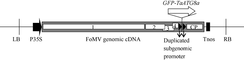
Bouton C, King R C, Chen H, Azhakanandam K, Bieri S, Hammond-Kosack K E, Kanyuka K. Foxtail mosaic virus: a viral vector for protein expression in cereals. Plant Physiol, 2018, 177:1352-1367.
doi: 10.1104/pp.17.01679
[6] Dommes A B, Gross T, Herbert D B, Kivivirta K I, Becker A. Virus-induced gene silencing: empowering genetics in non-model organisms. J Exp Bot, 2019, 70:757-770.
doi: 10.1093/jxb/ery411 pmid: 30452695
[7] Kant R, Dasgupta I. Gene silencing approaches through virus-based vectors: speeding up functional genomics in monocots. Plant Mol Biol, 2019, 100:3-18.
doi: 10.1007/s11103-019-00854-6
[8] Lee W S, Hammond-Kosack K E, Kanyuka K. Barley stripe mosaic virus-mediated tools for investigating gene function in cereal plants and their pathogens: virus-induced gene silencing, host- mediated gene silencing, and virus-mediated over-expression of heterologous protein. Plant Physiol, 2012, 160:582-590.
doi: 10.1104/pp.112.203489
[9] Liu N, Xie K, Jia Q, Zhao J, Chen T, Li H, Wei X, Diao X, Hong Y, Liu Y. Foxtail mosaic virus-induced gene silencing in monocot plants. Plant Physiol, 2016, 171:1801-1807.
doi: 10.1104/pp.16.00010 pmid: 27225900
[10] Mei Y, Beernink B M, Ellison E E, Konečná E, Neelakandan A K, Voytas D F, Whitham S A. Protein expression and gene editing in monocots using foxtail mosaic virus vectors. Plant Direct, 2019, 3:e00181.
[11] Li F, Vierstra R D. Autophagy: a multifaceted intracellular system for bulk and selective recycling. Trends Plant Sci, 2012, 17:526-537.
doi: 10.1016/j.tplants.2012.05.006
[12] Bassham D C. Function and regulation of macroautophagy in plants. Biochim Biophys Acta, 2009, 9:1397-1403.
[13] Nakatogawa H, Ichimura Y, Ohsumi Y. Atg8, a ubiquitin-like protein required for autophagosome formation, mediates membrane tethering and hemifusion. Cell, 2007, 130:165-178.
pmid: 17632063
[14] Izumi M, Hidema J, Wada S, Kondo E, Kurusu T, Kuchitsu K, Makino A, Ishida H. Establishment of monitoring methods for autophagy in rice reveals autophagic recycling of chloroplasts and root plastids during energy limitation. Plant Physiol, 2015, 167:1307-1320.
[15] Merkulova E A, Guiboileau A, Naya L, Masclaux-Daubresse C, Yoshimoto K. Assessment and optimization of autophagy monitoring methods in Arabidopsis roots indicate direct fusion of autophagosomes with vacuoles. Plant Cell Physiol, 2014, 55:715-726.
doi: 10.1093/pcp/pcu041
[16] Pei D, Zhang W, Sun H, Wei X J, Yue J Y, Wang H Z. Identification of autophagy-related genes ATG4 and ATG8 from wheat ( Triticum aestivum L.) and profiling of their expression patterns responding to biotic and abiotic stresses. Plant Cell Rep, 2014, 33:1697-1710.
doi: 10.1007/s00299-014-1648-x
[17] Li K X, Liu Y N, Yu B J, Yang W W, Yue J Y, Wang H Z. Monitoring autophagy in wheat living cells by visualization of fluorescence protein-tagged ATG8. Plant Cell Tissue Organ Cult, 2018, 134:481-489.
doi: 10.1007/s11240-018-1437-2
[1] ZHANG Wei,SUN Hong,WEI Xiao-Jing,XING Li-Ping,WANG Hua-Zhong. Cloning of Autophagy-Related Genes, ATG10s, in Wheat and Their Expression Characteristics Induced by Blumeria graminis f. sp. tritici [J]. Acta Agron Sin, 2014, 40(08): 1392-1402.
Viewed
Full text


Abstract

Cited

  Shared   
  Discussed   
No Suggested Reading articles found!